What is Thoracoscopy / Pleuroscopy?
Thoracoscopy, also known as medical pleuroscopy, is a safe, minimally invasive procedure used to examine the pleural space (the lining around the lungs), drain fluid, and take tissue samples. It is usually performed using local anesthesia and conscious sedation, avoiding the need for general surgery. A thin video-assisted thoracoscope is inserted through a small incision in the chest wall to inspect and biopsy the pleural cavity directly.
INDICATIONS
When Is It Recommended?
- Unexplained pleural effusion, meaning fluid build up in the pleural space, requiring proper pleural effusion treatment
- Suspected pleural malignancy, including mesothelioma or metastatic cancer spread to the pleura, needing accurate lung cancer diagnosis
- Pleural thickening or nodules seen on CT scan or ultrasound
- Pleural infections, such as empyema or tuberculosis related pleural disease, confirmed through tuberculosis diagnosis
Other Important Indications
- Recurrent fluid collection despite repeated needle drainage, often needing better lung fluid drainage
- Diagnosing the cause of persistent breathlessness or chest discomfort linked to fluid around the lungs, supported by proper chest fluid diagnosis
- Follow up evaluation after inconclusive fluid analysis or needle biopsy
Who Should Consider This Procedure?
Thoracoscopy and pleuroscopy services available in Navi Mumbai are generally recommended for patients who have fluid around the lungs that keeps coming back after drainage, a CT scan or ultrasound showing thickened or nodular pleura, suspected cancer spread to the chest lining, ongoing infection in the pleural space unresponsive to standard treatment, or symptoms like chest heaviness, breathlessness, or dry cough with an unclear cause. A pulmonologist or chest specialist typically assesses imaging, symptoms, and overall health before recommending this procedure as the right next step.
Why This Procedure Is Preferred
- Avoids the need for a larger open surgical approach
- Provides direct visual inspection of the pleura, improving diagnostic accuracy
- Allows fluid drainage and tissue biopsy in the same sitting, combining pleural biopsy with treatment
- Enables same session treatment, such as talc pleurodesis, to prevent fluid recurrence
- Performed under local anesthesia and sedation in most cases, so recovery is quicker
- Reduces the number of repeat procedures needed for diagnosis and treatment
- Widely available as a day care procedure at leading chest and lung care centers across Navi Mumbai
Procedure & Technique
Preparation & Aftercare
- Pre-procedure evaluation: Blood tests for coagulation, along with recent chest imaging, are usually reviewed before the procedure.
- Fasting: Patients are asked to fast for a few hours as per standard protocol before sedation.
- Duration: The procedure usually takes about 45 to 60 minutes, depending on the findings and treatment given.
- In-hospital stay: A short hospital stay is common, and a temporary chest tube may be left in place briefly to drain any remaining fluid.
- Monitoring: A chest radiograph is usually taken after the procedure to rule out complications such as pneumothorax.
- Discomfort: Mild pain or shoulder discomfort is possible for a day or two, usually managed with simple pain relief medication.
- Follow up: A follow up visit with the treating pulmonologist or chest specialist is usually scheduled to discuss biopsy results and plan further treatment if needed.
Risks / Complications
- Bleeding at the incision site or within the chest, usually minor
- Infection, which is uncommon with proper aftercare
- Lung injury or partial lung collapse, known as pneumothorax
- Persistent air leak in rare cases, needing a slightly longer chest tube duration
- Pain or soreness around the incision site
- Rare reaction to sedative medication, closely monitored throughout
How Results Are Used
Fluid and tissue samples collected during the pleural biopsy procedure are sent for detailed laboratory testing, including cytology, histopathology, and microbiology. Based on these results, the diagnosis is confirmed, whether it points to infection, tuberculosis diagnosis, or lung cancer diagnosis, and the next steps are planned, such as cancer treatment, TB medication, infection management, or further tests if the initial findings are unclear.
Conclusion
Thoracoscopy Navi Mumbai and pleuroscopy Navi Mumbai procedures provide a safe and accurate way to diagnose and manage pleural diseases with minimal surgical intervention. The procedure combines diagnosis and treatment in a single sitting, offering quicker recovery compared to traditional open surgery, and remains one of the most reliable methods for pleural effusion treatment in Navi Mumbai and evaluation of pleural infections or suspected pleural cancer. This procedure is performed by Dr. Vinod B. Chavhan, a pulmonologist and chest specialist based in Navi Mumbai.
Common Questions Patients May Have
About the Consultant
Dr. Vinod B. Chavhan is a pulmonologist and chest specialist based in Navi Mumbai, providing diagnosis and treatment for pleural effusion, pleural infections, tuberculosis, and lung related conditions, including thoracoscopy and pleuroscopy procedures.
Clinic Locations
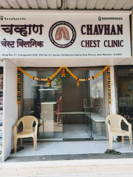
Chavhan Chest Clinic - Panvel
📍 Shop No-21, Indrsprasht CHS, Plot No-1, Sector-10, Khanda Colony, New Panvel East, Navi Mumbai, Maharashtra 410206
Phone: +91 9004985020
Get Location
Chavhan Chest Clinic - Kharghar
📍 Shop No 312-313, 3rd Floor, Pacific Building, Shilp Chowk, Sector 13, Kharghar, Navi Mumbai, Maharashtra 410210
Phone: +91 9820676822
Get Location
Kokilaben Dhirubhai Ambani Hospital
📍 Thane-Belapur Road, Kopar Khairane, Navi Mumbai, Maharashtra 400710
Phone: +91 9004985020
Get Location
Sanjivani Speciality Clinic - Vashi
📍 Office No. 706, JK Chambers Building, Sector 17, Vashi, Navi Mumbai, Maharashtra 400703
Phone: +91 9820676822
Get Location