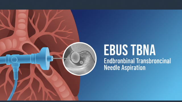
What is EBUS TBNA?
EBUS-TBNA (Endobronchial Ultrasound-guided Transbronchial Needle Aspiration) is a safe, minimally invasive bronchoscopic procedure that combines real-time ultrasound imaging with fine-needle aspiration through the airway wall. It allows the doctor to sample lymph nodes or masses located next to the airways (mediastinal and hilar lymph nodes) without the need for open surgery. This advanced bronchoscopy technique is widely used for lung cancer diagnosis, lung cancer staging, tuberculosis (TB) detection, sarcoidosis, and other inflammatory or infectious chest conditions.
If you are searching for an experienced pulmonologist near me in Navi Mumbai for lung biopsy or chest lymph node evaluation, Dr. Vinod B. Chavhan offers advanced, accurate, and minimally invasive diagnostic care.
Purpose / Indications
- Staging of lung cancer by sampling mediastinal/hilar lymph nodes
- Diagnosis of mediastinal lymphadenopathy (sarcoidosis, lymphoma, TB/tuberculosis)
- Biopsy of peribronchial lesions and lung masses
- Evaluation of unexplained enlarged chest lymph nodes seen on CT scan or PET scan
- Diagnosing chronic cough, unexplained chest lymph node swelling, or suspected lung infection
- Assessing recurrence of cancer after chemotherapy or radiation treatment
- Investigating fever of unknown origin linked to chest lymph nodes
- Evaluating patients with abnormal chest X-ray findings that need tissue confirmation
Who Should Consider EBUS TBNA?
- Enlarged lymph nodes in the chest detected on a CT or PET scan
- A suspicious lung mass that needs tissue diagnosis
- Known or suspected lung cancer that requires staging before treatment planning
- Symptoms such as persistent cough, unexplained weight loss, night sweats, or breathlessness
- A history of TB exposure with unclear chest imaging findings
- Suspected sarcoidosis or other granulomatous lung disease
Your pulmonologist will review your symptoms, scan reports, and overall health before recommending EBUS TBNA as the most suitable diagnostic option.
Why Choose This Procedure?
- Avoids invasive surgical mediastinoscopy
- High diagnostic accuracy with lower risk and faster recovery than surgery
- Real-time ultrasound image guidance increases precision of tissue sampling
- Minimal discomfort compared to traditional surgical lung biopsy
- Can reach lymph node stations that are difficult to access with standard bronchoscopy
- Reduces hospital stay, since most patients are treated as day care cases
- Trusted diagnostic tool used by leading chest specialists and pulmonologists worldwide
- Allows rapid on site evaluation of samples in many centers, speeding up diagnosis
How the Procedure Works
EBUS-TBNA uses an ultrasound-guided bronchoscope to collect tissue or fluid samples from lymph nodes in the chest, helping doctors accurately diagnose lung cancer, tuberculosis, and inflammatory lung diseases.
Scope Insertion
A thin, flexible tube fitted with a camera and ultrasound probe is gently passed through the mouth or nose into the windpipe (trachea).
Real-Time Ultrasound View
The doctor uses sound waves (ultrasound) to clearly visualize lymph nodes and masses located just outside the airway walls, including their size, shape, and blood flow pattern.
Needle Sample
A fine needle is passed through the scope to collect cells or tissue for laboratory and pathology testing. Multiple passes from different angles may be taken to ensure an adequate sample.
Sample Handling & Testing
Collected samples are sent for cytology, histopathology, microbiology, or molecular testing, depending on the suspected condition.
Preparation & What to Expect
- Fasting: Patients are advised to fast for a few hours before the procedure, usually 6 to 8 hours for food and fewer hours for clear liquids.
- Medical history review: You will be asked about current medications, allergies, bleeding disorders, and blood thinners, since some medicines may need to be paused before the procedure.
- Blood tests: Basic blood tests may be done beforehand to check clotting function and overall fitness for sedation.
- Sedation: The procedure is performed under moderate sedation or local anesthesia (occasionally general anesthesia), so you remain comfortable throughout.
- Duration: The entire process usually takes about 30 to 60 minutes, depending on the number of lymph node stations sampled.
- Monitoring: IV access is established, and vital signs such as oxygen levels, heart rate, and blood pressure are monitored throughout the procedure.
Recovery & Aftercare
- Mild sore throat, occasional cough, or minimal bleeding may occur. These are normal and temporary.
- You will rest in the recovery area until the throat numbness wears off, usually within an hour or two.
- Most patients are discharged the same day and can resume normal activities within 24 hours.
- You should avoid eating or drinking until the throat sensation fully returns, to prevent accidental choking.
- Someone should accompany you home, since sedation can affect alertness and reaction time for several hours.
- Report to your doctor immediately if you notice heavy bleeding, high fever, chest pain, or severe breathlessness after the procedure.
Risks / Complications
- Bleeding (usually minor and self limiting)
- Infection (rare)
- Pneumothorax, or collapsed lung (rare)
- Transient hypoxia, meaning a temporary drop in oxygen levels
- Sore throat or hoarseness lasting a day or two
- Reaction to sedative medication, which is uncommon and closely monitored
How Results Are Used
Once the sample is collected, it is examined by a pathologist under a microscope and, if needed, tested further for infections or specific cancer markers. Based on the findings, your pulmonologist will discuss the diagnosis with you and plan the next steps, which may include starting cancer treatment, TB medication, anti-inflammatory therapy, or further tests if the initial sample is inconclusive.
Conclusion
EBUS-TBNA offers a safe, accurate, and minimally invasive way to diagnose lung cancer, tuberculosis, sarcoidosis, and other chest conditions, with minimal discomfort and a much quicker recovery compared to surgical biopsy methods. If you are looking for expert EBUS TBNA treatment in Navi Mumbai, Dr. Vinod B. Chavhan provides advanced diagnostic bronchoscopy services with real time ultrasound guidance for accurate lymph node and lung mass evaluation.
Common Questions Patients May Have
Are you or a loved one scheduled for an EBUS-TBNA?
Do you have questions about preparation, sedation, or potential risks?
Clinic Locations
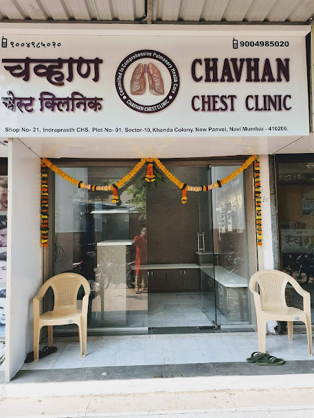
Chavhan Chest Clinic - Panvel
📍 Shop No-21, Indrsprasht CHS, Plot No-1, Sector-10, Khanda Colony, New Panvel East, Navi Mumbai, Maharashtra 410206
Phone: +91 9004985020
Get Location
Chavhan Chest Clinic - Kharghar
📍 Shop No 312-313, 3rd Floor, Pacific Building, Shilp Chowk, Sector 13, Kharghar, Navi Mumbai, Maharashtra 410210
Phone: +91 9820676822
Get Location
Kokilaben Dhirubhai Ambani Hospital
📍 Thane-Belapur Road, Kopar Khairane, Navi Mumbai, Maharashtra 400710
Phone: +91 9004985020
Get Location
Sanjivani Speciality Clinic - Vashi
📍 Office No. 706, JK Chambers Building, Sector 17, Vashi, Navi Mumbai, Maharashtra 400703
Phone: +91 9820676822
Get Location