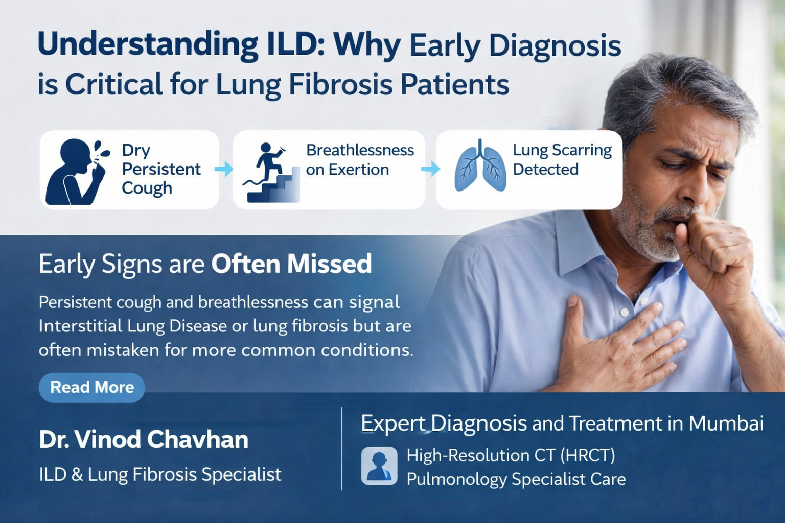
Understanding ILD: Why Early Diagnosis is Critical for Lung Fibrosis Patients
If you have been coughing for weeks and no one seems to know why, you are not imagining it. A persistent dry cough that just will not go away, combined with shortness of breath while climbing stairs or doing routine tasks, are two of the most commonly ignored warning signs of a serious lung condition. Most patients spend months visiting general physicians before anyone mentions the words Interstitial Lung Disease symptoms or lung fibrosis. Many of them only get answers after finally reaching Pulmonology Specialist in Mumbai who recognize the pattern immediately. This blog is for those patients. The ones sitting with a report they do not fully understand, or the ones who have been told “your lungs show some changes” without a clear explanation of what that actually means.
What Is Interstitial Lung Disease, and Why Does It Get Missed?
Interstitial Lung Disease, or ILD, is not a single disease. It covers more than 200 different conditions that cause scarring or inflammation inside the lung tissue. The lungs lose their ability to stretch and fill properly, which is why breathlessness (Dyspnea) is such a consistent complaint.
The reason ILD gets missed so often is that its early symptoms look exactly like something more common. A dry cough gets treated as allergies. Breathlessness gets blamed on fitness levels or anxiety. By the time a patient sees a specialist, the scarring may already be progressing.
Three conditions frequently confused with each other are:
ILD (Interstitial Lung Disease): The broad category. Scarring and inflammation in the interstitium, the tissue that surrounds the air sacs of the lung. Causes range from autoimmune disease to dust exposure to unknown triggers.
HP (Hypersensitivity Pneumonitis): An immune reaction triggered by inhaling organic dust, mold, bird droppings, or certain chemicals. The lungs are reacting to something in the environment. Find the trigger and remove it early, and the lung has a real chance to recover. Miss it, and it becomes chronic fibrosis.
IPF (Idiopathic Pulmonary Fibrosis): The most severe form. Idiopathic means the cause is unknown. The scarring in IPF is progressive and irreversible, which is exactly why lung fibrosis treatment 2026 focuses so heavily on slowing the disease rather than reversing it. Early diagnosis here is not just helpful. It is the difference between preserving lung function and losing it permanently.
Recognizing the Early Warning Signs
The challenge with ILD is that no single symptom points directly to the disease. Patients often describe a gradual onset over months, which is easy to rationalize away.
The most common early Interstitial Lung Disease symptoms include:
Dry, persistent cough that does not respond to standard cough medications or antihistamines. Unlike a productive cough from infection, this one produces little or no mucus and tends to worsen with physical activity or lying down.
Progressive breathlessness (Dyspnea) that starts with exertion and slowly creeps into rest. Many patients first notice it when walking up a flight of stairs, carrying groceries, or talking for extended periods.
Fatigue and reduced exercise tolerance. This is often attributed to aging or poor fitness, but in ILD it connects directly to reduced oxygen exchange in scarred lung tissue.
Clubbing of the fingers. In some ILD subtypes, particularly IPF, the fingertips widen and the nails curve downward. This is a late sign but an important physical finding.
Crackling sounds in the lungs. A doctor listening with a stethoscope may detect a distinctive sound, often described as Velcro tearing, at the base of the lungs. Stiff, scarred tissue opening with each breath causes it.
If you have two or more of these, the next step is not another course of antibiotics. It is a specialist evaluation.
Risk Factors That Increase ILD Likelihood
ILD does not develop randomly. Certain profiles carry a meaningfully higher risk.
Occupational exposure is one of the most underappreciated risk factors. Construction workers, farmers, hairdressers, and those exposed to silica dust, asbestos, bird feathers, or mold spores over years are at elevated risk for HP and other occupational ILDs.
Autoimmune diseases including Rheumatoid Arthritis, Scleroderma, Lupus, and Sjogren’s Syndrome frequently involve the lungs. Patients with these diagnoses should have periodic pulmonary assessments even before respiratory symptoms appear.
Smoking history does not cause IPF directly, but it is a recognized risk factor. Smokers with unexplained respiratory symptoms should not assume their cough is simply a smoker’s cough.
Age and sex. IPF specifically tends to affect men over 60, though ILD as a broader category affects both sexes across age groups.
Genetic predisposition. A family history of pulmonary fibrosis raises the likelihood, and some ILD subtypes have identifiable genetic mutations now being studied in lung fibrosis treatment 2026 research programs.
Why an HRCT Scan Is Not Optional
A standard chest X-ray will miss early ILD. X-rays are designed to catch pneumonia, fractures, and obvious masses. The fine, web-like scarring patterns of ILD require a different tool entirely.
An HRCT scan (High Resolution CT) gives the doctor a detailed, cross-sectional map of your lung tissue. It identifies the pattern of scarring, whether it sits at the bases of the lungs, whether it looks like honeycombing, and whether it matches a known ILD subtype. This distinction matters because HP and IPF are managed differently. Treating one like the other can accelerate damage.
HRCT findings that suggest ILD include:
Ground glass opacities: Hazy areas that suggest active inflammation. These can partially resolve with treatment in HP, making them an important prognostic marker.
Reticular pattern: A net-like pattern of fine lines across the lung tissue, common in early fibrosis.
Honeycombing: Clustered air-filled cysts, usually in the lower lung zones. This pattern in IPF indicates advanced fibrosis and drives treatment decisions under lung fibrosis treatment 2026 protocols.
Traction bronchiectasis: Airways pulled open and distorted by surrounding scar tissue.
Any best pulmonologist for ILD will tell you that an HRCT is the first real diagnostic step. If your doctor has not recommended one and your symptoms have persisted for more than eight weeks, ask for it directly.
When Bronchoscopy Becomes Necessary
An HRCT tells you what the scarring looks like. A bronchoscopy tells you what is happening at the cellular level. A thin, flexible tube is guided through the airway so the doctor can visually examine the airways and collect samples from deep inside the lung tissue.
Bronchoscopy with Bronchoalveolar Lavage (BAL) washes a small section of the lung and collects the fluid for analysis. This can reveal immune cell patterns that confirm HP, or rule out infection and malignancy, which can mimic ILD on imaging. In some cases, a transbronchial biopsy is taken to examine actual lung tissue.
A BAL result showing a high proportion of lymphocytes strongly supports a diagnosis of HP rather than IPF. This single distinction changes the entire management plan. HP may respond to steroids and antigen avoidance. IPF does not respond to steroids and instead requires antifibrotic medication.
Not every ILD patient needs a bronchoscopy. But when the HRCT is inconclusive, or when the clinical picture does not match the imaging, it becomes a critical next step. Skipping it to avoid the procedure can delay the correct diagnosis by months.
In selected cases where bronchoscopy is inconclusive, a surgical lung biopsy via Video-Assisted Thoracoscopic Surgery (VATS) may be recommended. This provides a larger tissue sample for definitive pathological diagnosis. The decision always involves weighing diagnostic certainty against procedure risk, a judgment call made by an experienced ILD specialist in Mumbai in close discussion with the patient.
Pulmonary Function Tests: Measuring What Imaging Cannot
Imaging shows the structural damage. Pulmonary Function Tests (PFTs) measure how much that damage has affected breathing mechanics. Together, they give a complete picture.
The most relevant PFT findings in ILD include:
Reduced FVC (Forced Vital Capacity): The total amount of air a person can forcibly exhale. In ILD, this is reduced because stiff lungs cannot fully expand.
Reduced DLCO (Diffusion Capacity): This measures how efficiently oxygen crosses from the lung air sacs into the bloodstream. Fibrosis creates a physical barrier that slows this transfer even before the patient feels significantly breathless.
Preserved or elevated FEV1/FVC ratio: Unlike obstructive diseases like COPD where this ratio drops, in ILD it often stays normal or rises. This pattern, called restrictive physiology, is one of the functional signatures of ILD.
Serial PFTs done every three to six months allow the specialist to track disease progression objectively. A decline in FVC of 10 percent or more over 12 months is a significant event that typically prompts treatment escalation under current lung fibrosis treatment 2026 guidelines.
Lung Elasticity and Why It Has to Be Protected Early
Healthy lung tissue has a natural elasticity. It expands with each breath and recoils to push air out. Fibrosis replaces that elastic tissue with stiff scar tissue. Once that happens, the lung cannot stretch or recoil properly, and breathing becomes a physical effort.
The goal of early intervention is not just symptom control. It is protecting whatever elasticity remains. Antifibrotic medications available under lung fibrosis treatment 2026 protocols can slow scar formation but they cannot undo damage that is already there. This is what makes timing so consequential.
Patients who receive the right diagnosis within the first six months of symptom onset generally retain significantly more lung function than those diagnosed after a year or more. That gap in outcomes is the real argument for moving quickly when symptoms appear.
Current Treatment Approaches for ILD and Lung Fibrosis
Treatment in ILD is not one-size-fits-all. The subtype of disease determines the strategy.
For IPF, the current standard of care involves antifibrotic medications. Two drugs, nintedanib and pirfenidone, are approved and shown to slow FVC decline. They do not reverse fibrosis, but they slow the rate at which the disease progresses. Lung fibrosis treatment 2026 research is exploring combination approaches, biomarker-guided therapy, and newer antifibrotic agents currently in trials.
For HP, the immediate priority is identifying and removing the causative antigen. If caught early, some patients show meaningful recovery of lung function after antigen removal alone. When inflammation persists, corticosteroids suppress the immune reaction. Chronic HP that has progressed to fibrosis is managed more like IPF with antifibrotic therapy.
For autoimmune-related ILD, treatment targets the underlying autoimmune disease with immunosuppressants such as mycophenolate mofetil or azathioprine, often in coordination with a rheumatologist.
Pulmonary rehabilitation is recommended across all ILD subtypes. Supervised exercise training, breathing techniques, and education about the disease improve functional capacity and quality of life even when the underlying fibrosis cannot be reversed.
Oxygen therapy becomes necessary when resting oxygen levels fall below a certain threshold. Long-term supplemental oxygen protects the heart and improves exercise tolerance.
Lung transplantation remains an option for carefully selected patients with end-stage disease who are otherwise fit for the procedure. Referral to a transplant center is a decision made jointly between the patient and the treating pulmonologist.
Pulmonology Specialists in Mumbai: What to Look For and Why It Matters
Mumbai has some of the country’s most experienced Pulmonology Specialist in Mumbai, but not all of them see ILD cases regularly. This distinction matters more than most patients realize.
ILD is a subspecialty within pulmonology. A chest physician who primarily manages asthma, COPD, and respiratory infections handles fundamentally different clinical problems. The diagnostic reasoning required for ILD, reading an HRCT pattern, interpreting BAL cell differentials, deciding between antifibrotic therapy and immunosuppression, is built through repeated exposure to complex lung cases over years. Pulmonology doctors in Mumbai who focus on ILD develop this pattern recognition in a way that generalists simply cannot replicate.
What separates an ILD-focused pulmonologist from a general chest physician in practice:
Familiarity with ILD subtypes. There are more than 200 causes of ILD. A specialist who sees these cases regularly knows the clinical presentations of HP, IPF, connective tissue disease-associated ILD, sarcoidosis, and rare subtypes like PLCH or LAM. This experience changes the diagnostic questions being asked from the very first appointment.
Ability to interpret HRCT in clinical context. An HRCT report from a radiologist describes findings. An ILD-experienced pulmonologist correlates those findings with the patient’s occupational history, autoimmune markers, and symptom timeline to arrive at a working diagnosis. Two patients with identical HRCT reports can have completely different underlying conditions.
Access to bronchoscopy and advanced diagnostic tools. Pulmonology Specialists in Mumbai operating in well-equipped centers can perform or coordinate BAL, transbronchial biopsy, and surgical biopsy when indicated, rather than relying on imaging alone.
Experience managing antifibrotic therapy. Nintedanib and pirfenidone have side effect profiles that require active management. Gastrointestinal side effects with pirfenidone and bleeding risk with nintedanib are practical challenges that an experienced specialist navigates without unnecessarily stopping effective therapy.
Multidisciplinary collaboration. Many ILD cases involve autoimmune overlap, which means the pulmonologist needs to coordinate with a rheumatologist. ILD specialist in Mumbai working within larger clinical settings are better positioned for this kind of integrated care.
For patients looking for a pulmonologist in Mumbai, the right questions to ask include: How many ILD patients does the physician see per month? Does the clinic have HRCT interpretation capability on-site? Is PFT testing available? Can bronchoscopy be performed and interpreted by the same team? These are not demanding questions. They are the basic requirements for competent ILD care.
Why Mumbai Patients Seek a Specialist Rather Than a General Physician
ILD is not managed with a prescription and a follow-up appointment. It requires an integrated approach involving HRCT interpretation, pulmonary function testing, bronchoscopy when indicated, and a treatment plan that accounts for the specific subtype of disease and the patient’s overall health.
This is why patients across Maharashtra look for an ILD specialist in Mumbai rather than relying on a general chest physician. The diagnostic nuances between HP and IPF require a physician who sees these cases regularly and knows what pattern of treatment response to expect.
For patients already diagnosed elsewhere, seeking a top ILD specialist for second opinion in Mumbai is a reasonable and often important step. Treatment decisions in ILD, particularly around starting antifibrotic therapy or immunosuppression, carry significant consequences. A second opinion from a specialist who works exclusively with complex lung conditions can confirm a diagnosis or catch something initially missed.
Second opinions in ILD frequently change the management plan. Reviews from multiple ILD referral centers show that second-opinion evaluations led to a change in diagnosis or treatment approach in nearly a third of cases. That is not a commentary on the quality of initial care. It reflects how genuinely complex this disease category is. Getting a second opinion is not about distrust. It is about certainty, and in a progressive disease, certainty matters.
Dr. Vinod Chavhan: ILD and Lung Fibrosis Care in Mumbai
Dr. Vinod Chavhan is a pulmonologist with focused expertise in managing Interstitial Lung Disease symptoms, lung fibrosis treatment 2026 protocols, and complex cases involving HP and IPF. His approach centers on early and accurate diagnosis through HRCT interpretation and bronchoscopy, followed by individualized treatment plans designed to preserve lung function and slow fibrosis progression.
Patients visiting his pulmonology clinic near me searches from across Mumbai and Navi Mumbai come with a wide range of presentations, from early unexplained dry cough to advanced fibrosis requiring antifibrotic therapy. His clinic handles the full diagnostic workup in one place, cutting the delays that come from coordinating between multiple facilities.
For patients managing costs alongside a serious diagnosis, affordable lung fibrosis care in Navi Mumbai is available through his practice without compromising the quality of diagnostic tools or treatment protocols.
When to Stop Waiting and Make an Appointment
These are the situations where you should contact a specialist without further delay:
You have had a dry cough for more than six to eight weeks with no clear cause. You feel breathless doing activities that did not previously tire you out. Your doctor has mentioned “ground glass changes” or “fibrosis” on a chest scan. You have already been diagnosed with ILD but have questions about your treatment plan. You have a history of autoimmune disease and are now developing respiratory symptoms. You work in an occupation involving dust, mold, chemicals, or birds and have any respiratory symptoms.
ILD does not pause while you wait for a convenient time to get checked. The window for protecting lung function is real, and it closes.